When we say “heartbeat” we don’t mean “fetal pole cardiac activity.” We mean “heartbeat.”
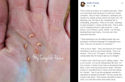
Recently a FB follower shared this post to our page:
 |
| (Click to enlarge) |
This is what an embryo at 6 weeks looks like. There is no real heart beat because it’s heart isn’t nearly complete – they’re heart “vibrations” (vibrations are caused my cellular activity where the heart WILL be. Meaning, yes, the title of the “heartbeat bill” is misleading, purposely). There is no brain, meaning no pain receptors. It does not feel pain. This is what you’re stripping women’s right away for. I, your sisters, your mothers, aunts, friends – we all have beating hearts and brains. Our lives are more important than this.
**Stop listening to pro life talking heads that use purposely emotional language to manipulate your view. They are not doctors or scientists.**
•This is not a “baby”. They use pictures of 6 month old babies to pull on your heart strings. This is an embryo. This is not “10 fingers, 10 toes” babbling cooing baby they’re trying to get you to imagine.
The post is certainly right that this image is not of a “baby.” The image is actually from Etsy, described as “Baby Memorial/Honor Sculpture.” The tiny figures pictured are clay sculptures which the seller says are “for those who have experienced the loss of a pregnancy during the first trimester and are searching for a tangible keepsake to honor their precious Angel.” The Etsy page includes reviews from mothers describing how much it means to them to have a way to mark their grief and loss. How ironic that the OP uses art specifically meant to help people value and mourn prenatal life to instead deride those very viewpoints–and all while claiming to be representing science. It’s kind of amazing.
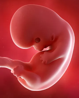
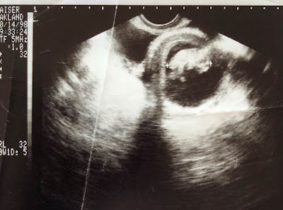
Here’s an image of an embryo around 6 weeks post-fertilization (or 8 weeks LMP – after the beginning of the last menstrual period) courtesy of The Bump:
The Bump’s use of the word “heartbeat” is representative of not only many prenatal websites but also descriptions medical professionals give pregnant women during routine prenatal care. Using “heartbeat” to describe embryonic activity at this stage is neither new nor unique to anti-abortion advocates.
Some pro-choice people argue that when medical professionals say “heartbeat” in these contexts, they’re just using layman’s language with their patients, just as an OBGYN might say “baby” when talking to a woman with a wanted pregnancy. That doesn’t make “baby” a medical or technical term.
But “heartbeat” is appropriate both for the layman and as a medical description. As The Developing Human by Moore et al (10th Edition, 2013) explains in “Chapter 13: Cardiovascular System”:
The cardiovascular system is the first major system to function in the embryo. The primordial heart and vascular system appear in the middle of the third week (Fig. 13-1). This precocious cardiac development occurs because the rapidly growing embryo can no longer satisfy its nutritional and oxygen requirements by diffusion alone. Consequently, there is a need for an efficient method of acquiring oxygen and nutrients from the maternal blood and disposing of carbon dioxide and waste products.
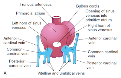
In other words the embryonic heart exchanges oxygen and carbon dioxide even before it fully develops into the more complex heart we’re familiar with. Those insisting we say “fetal pole cardiac activity” instead of “heartbeat” or describing the embryonic heart as just “electrically induced flickering” or–more ridiculously–“vibrations” try to imply that the four chambered heart doesn’t happen until months later; that’s completely incorrect. Here’s a diagram from Moore et al of the heart at 35 days (approximately 5 weeks post-fertilization):
 |
| (Click to enlarge) |
At this point the embryonic heart already has four chambers. It’s reductive to describe this development as no more than “pulsing cells.”
Additionally, by 4 weeks the embryo has three paired veins draining into the heart: vitelline veins return poorly oxygenated blood from the umbilical vessel, umbilical veins carry well-oxygenated blood from the chorionic sac, and cardinal veins return poorly oxygenated blood from the embryo’s body to the heart. Here is an illustration from figure 13-5 of Moore of the heart at 24 days postfertilization:
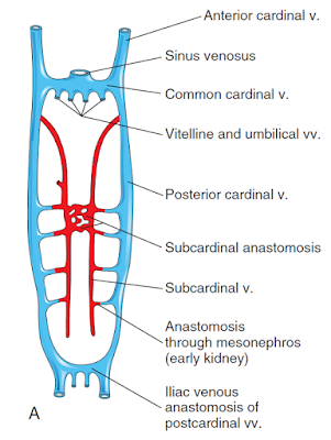
Here are the veins illustrated at 6 weeks:
This image is from Figure 13-4, the caption for which states, “Initially, three systems of veins are present: the umbilical veins from the chorion, vitelline veins from the umbilical vesicle, and cardinal veins from the body of the embryos.”
More from Moore:
- “The heart begins to beat at 22 to 23 days (Fig. 13-2).”
- “Blood flow begins during the fourth week, and heartbeats can be visualized by Doppler ultrasonography (Fig. 13-3).”
- “The initial contractions of the heart are of myogenic origin (in or starting from muscle). … At first, circulation through the primordial heart is an ebb-and-flow type; however, by the end of the fourth week, coordinated contractions of the heart result in unidirectional flow.”
- “Partitioning of the AV canal, primordial atrium, ventricle, and outflow tract begins during the middle of the fourth week.”
Post-publication update: I’ve seen so many stories now purporting to scientifically explain away the embryonic heart, I’m just going to start collecting them here:
- ‘Heartbeat’ Bills Get The Science of Fetal Heartbeats All Wrong, Wired, 5/14/19
- Is a ‘Fetal Heartbeat’ Really a Heartbeat at 6 Weeks? LiveScience, 5/17/19
- Embryos Don’t Have Hearts, The Cut, 5/24/19
- Doctors’ organization: calling abortion bans ‘fetal heartbeat bills’ is misleading, The Guardian, 6/5/19


Leave a Reply
Want to join the discussion?Feel free to contribute!