Embryonic Hearts
Intro
In response to states passing heartbeat bills (legislation that prohibits most abortions once an embryo’s heartbeat is detectable), abortion advocates claim embryos at early stages of pregnancy don’t have hearts or heartbeats. This is a myth. Abortion advocates sometimes use opaque descriptions such as “group of cells with electrical activity.” Such descriptions aren’t technically incorrect, but they obscure more than they reveal.
Please note that there are two methods of measuring gestational age. The medical community commonly measures based on the first day of the woman’s Last Menstrual Period (LMP), but in other instances gestational age is measured in days or weeks post-fertilization. On average, fertilization occurs about two weeks after LMP, so there is a two week difference in these two measurement methods. For example, 4 weeks post-fertilization is 6 weeks LMP.
By 6-7 weeks LMP (4-5 weeks post-fertilization), the embryo has:
- a chambered heart
- using coordinated muscle contractions
- to unidirectionally pump blood
- through veins
- to exchange oxygen and carbon dioxide.
Below are scientific sources establishing as much. We also list many of the below sources on Twitter here.
Development of the heart: Morphogenesis, growth, and molecular regulation of differentiation (link)
Figure 1.14 of the linked PDF contains scanning electron microscope images of the human embryonic heart. The upper right image from the figure is at 33 days post-fertilization (about 6.5 weeks LMP) and labels the four chambers (right and left atria, right and left ventricle).
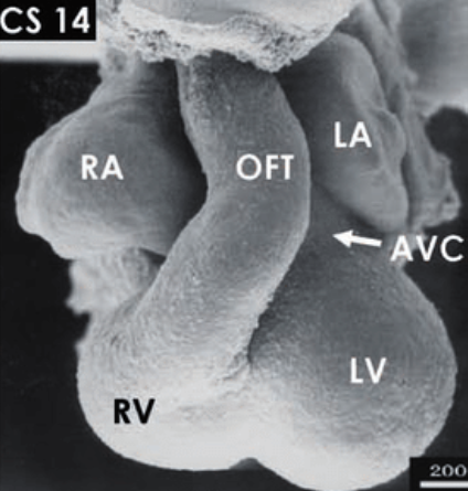
RV & LV = right and left ventricle;
OFT = outflow tract;
AVC = atrioventricular canal;
CS 14 = Carnegie Stage 14 (around 33 days postfertilization)
Just prior to Figure 1.14 is the section titled “Chamber Formation and Ventricular Septation,” which explains:
The electrical configuration of the embryonic heart consisting of alternating slowly and rapidly contracting segments, as described below, allows the early chamber-forming heart to produce coordinated atrial and ventricular contractions effectively propelling blood forward.
“Development of the Heart: Morphogenesis, Growth, and Molecular Regulation of Differentitation,” Sizarov, Baldwin, Srivastava, & Moorman, February 2016
Stages of Development of the Fetus (link)
This is a passage from the Merck Manual, one of the oldest continuously published English language medical textbooks. Under the subheading “Development of the Embryo,” the manual explains:
This [embryonic] stage characterized by the formation of most internal organs and external body structures. The heart and major blood vessels develop early, at about 16 days after fertilization. The heart begins to pump fluid and then blood through the blood vessels at about 5 weeks (3 weeks after fertilization).
“Stages of Development of the Fetus,” Merck Manual, Raul Artal-Mittelmark, revised Sept 2024
Anatomy and Physiology, an OpenStax online textbook; Chapter 19.5: Development of the Heart (link)
The lower left diagram in Figure 19.36 depicts the heart at 28 days post-fertilization (6 weeks LMP), including the four chambers with atrium and ventricle labeled.
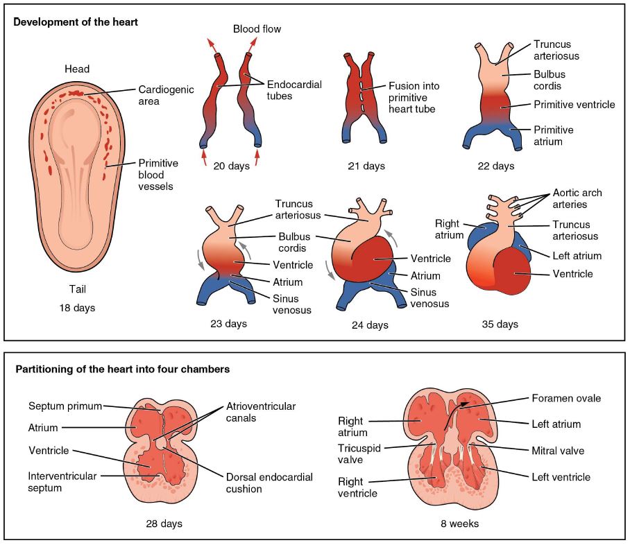
The chapter explains:
The five regions of the primitive heart tube develop into recognizable structures in a fully developed heart. … As the primitive heart tube elongates, it begins to fold within the pericardium, eventually forming an S shape, which places the chambers and major vessels into an alignment similar to the adult heart. This process occurs between days 23 and 28. The remainder of the heart development pattern includes development of septa and valves, and remodeling of the actual chambers. Partitioning of the atria and ventricles by the interatrial septum, interventricular septum, and atrioventricular septum is complete by the end of the fifth week [7 weeks LMP].
“Development of the Heart,” Betts et al, OpenStax, April 2013
The Transitional Heart: From Early Embryonic and Fetal Development to Neonatal Life (link)
This is an article published in the journal Fetal Diagnosis and Therapy by Tan & Lewandowski in 2019. In this article, the authors refer to gestational weeks, which are equivalent to LMP; in other words if the article says “gestational week 7” it refers to 7 weeks LMP. From the article’s introduction:
The human heart is one of the first organs to form and function during embryogenesis. By the end of gestational week 3, passive oxygen diffusion becomes insufficient to support metabolism of the developing embryo, and thus the fetal heart becomes vital for oxygen and nutrient distribution. The initiation of the first heart beat via the primitive heart tube begins at gestational day 22, followed by active fetal blood circulation by the end of week 4. … This eventually leads to the formation of the 4-chambered heart by gestational week 7.
“The Transitional Heart: From Early Embryonic and Fetal Development to Neonatal Life,” Tan & Lewandowski, Fetal Diagnosis and Therapy, 2020;47:373-386 September 2019
In Figure 2 part b, the lower right diagram depicts the human heart at gestational week 7:
PA = pulmonary artery
RA & LA = right and left atria
RV & LV = right and left ventricles
EPC = epicardium
IVC = inferior vena cavas
The figure’s caption states “The 4 chambers form by the end of week 7.”
The Developing Human: Clinically Oriented Embryology; Chapter 13: Cardiovascular System (link)
This is an embryology textbook for medical students written by Keith Moore, TVN Persaud, and Mark Torchia. The following information is from the 10th edition (published in 2013). Here is the first paragraph from Chapter 13: Cardiovascular System:
The cardiovascular system is the first major system to function in the embryo. The primordial heart and vascular system appear in the middle of the third week. This precocious cardiac development occurs because the rapidly growing embryo can no longer satisfy its nutritional and oxygen requirements by diffusion alone. Consequently, there is a need for an efficient method of acquiring oxygen and nutrients from the maternal blood and disposing of carbon dioxide and waste products.
The Developing Human, Moore, Persaud, & Torchia, 2013
In Figure 13-12 part D the diagram depicts the human heart at 35 days (5 weeks post-fertilization, 7 weeks LMP):
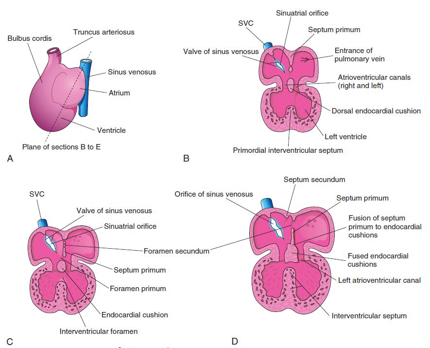
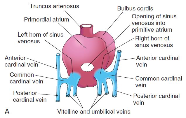
The text explains that by 24 days post-fertilization (about 5.5 weeks LMP), the embryo has three pairs of veins draining into the heart: (1) the vitelline veins return poorly oxygenated blood from the umbilical vessel, (2) umbilical veins carry well-oxygenated blood from the chorionic sac, and (3) cardinal veins return poorly oxygenated blood from the embryo’s body to the heart. Figure 13-5 part A is an illustration showing these veins:
Additional excerpts from the chapter:
The heart begins to beat at 22 to 23 days. Blood flow begins during the fourth week, and heartbeats can be visualized by Doppler ultrasonography.
The Developing Human, Moore, Persaud, & Torchia, 2013
The initial contractions of the heart are of myogenic origin (in or starting from muscle). … At first, circulation through the primordial heart is an ebb-and-flow type; however, by the end of the fourth week, coordinated contractions of the heart result in unidirectional flow.
The Developing Human, Moore, Persaud, & Torchia, 2013
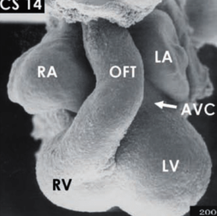
Formation of the building plan of the human heart (link)
This is an article published in the journal Circulation by Sizarov et al in 2011. In this research they examined the hearts of embryos collected after medically induced abortions. Here is Figure 5 (note row A, which is Carnegie Stage 10, has been omitted).
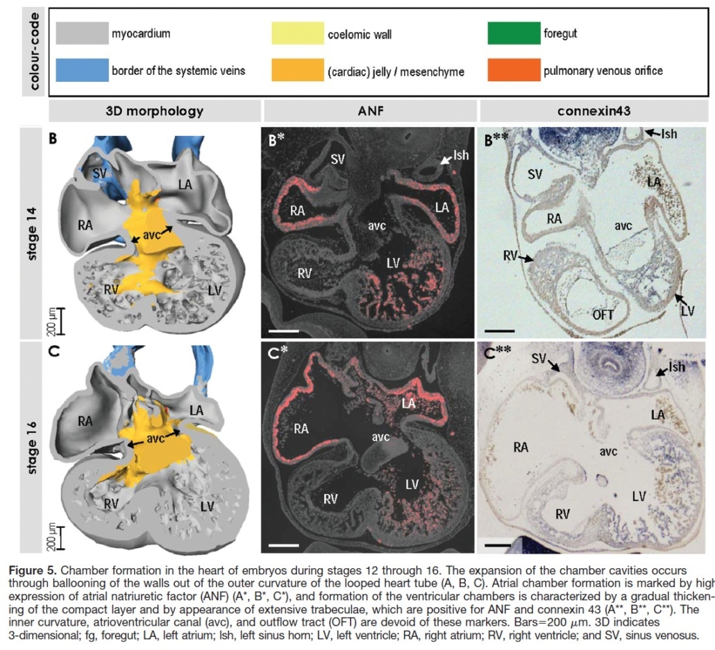
This figure shows the human embryonic heart at Carnegie Stage 14 (33 days postfertilization, or approximately 6.5 weeks LMP) and Carnegie Stage 16 (37-40 days postfertilization, or approximately 7 – 7.5 weeks LMP). Again RA & LA = right and left atria; RV & LV = right and left ventricle; and AVC = atrioventricular canal.
Dynamic transcriptome profiling toward understanding the development of the human embryonic heart during different Carnegie stages (link)
Article published in the Federation of European Biochemical Societies, Sept 2020
The development of electrical activities dramatically regulates the rhythm of the heart, which is consistent with the embryonic heart starting to beat at CS12 [between 6 – 6.5 weeks LMP].
“Dynamic transcriptome profiling toward understanding the development of the human embryonic heart during different Carnegie stages” Zhuo Meng et al September 2020
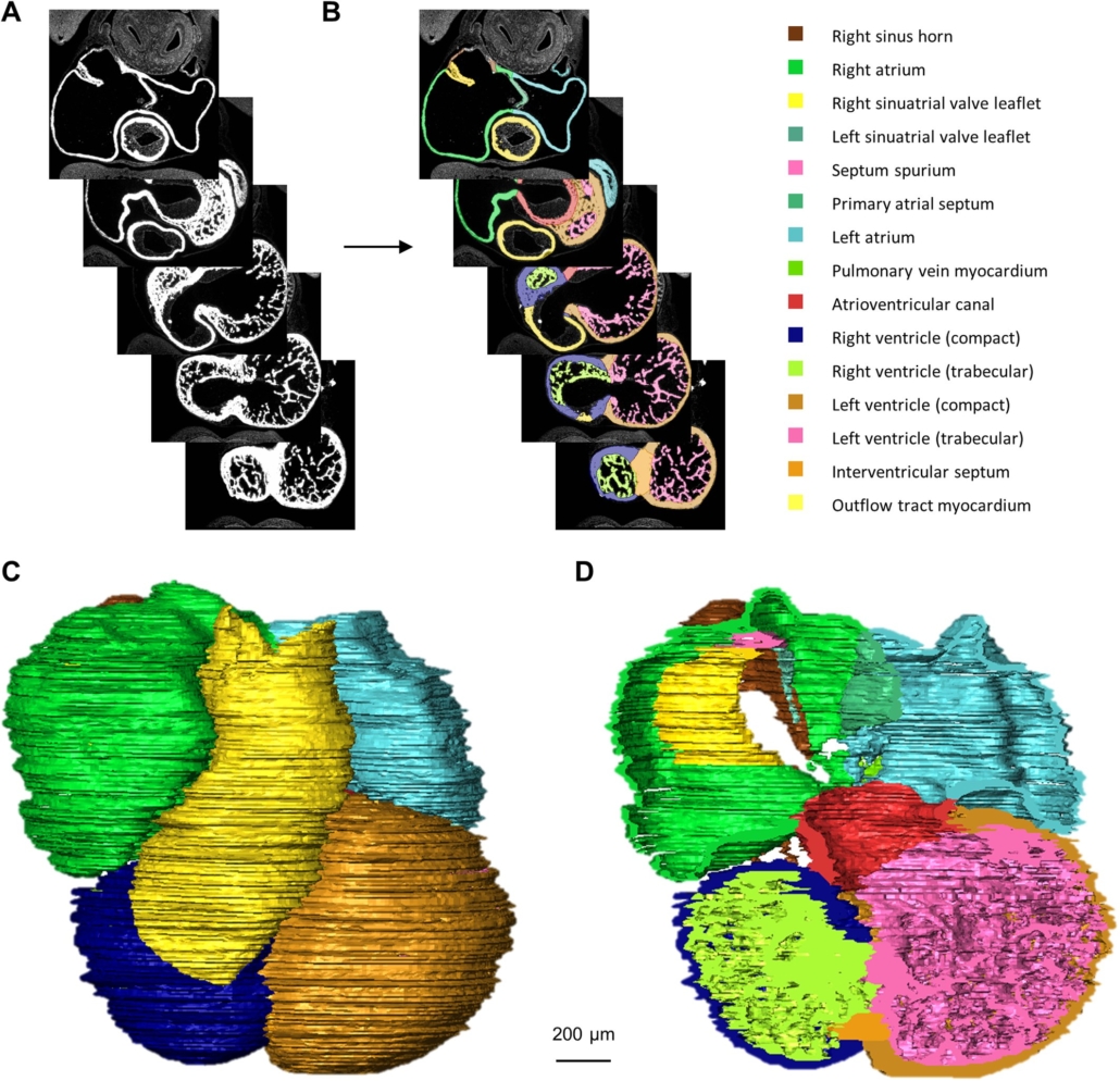
Quantified growth of the human embryonic heart (link)
This article was published by Faber et al in 2021 in Biology Open, an open access journal that publishes rigorously conducted high-quality biological and biomedical research. Figure 1 shows the chambers and other structures of the embryonic heart at Carnegie Stage 14, about 6.5 weeks LMP.